


Liam On Social

Meet Liam
-
![]()
The Odds...
That you’ll be struck by lightning this year is 1 in 500,000. The odds of a child being diagnosed with Juvenile Myositis are the same.
Meet our son Liam.
-
![]()
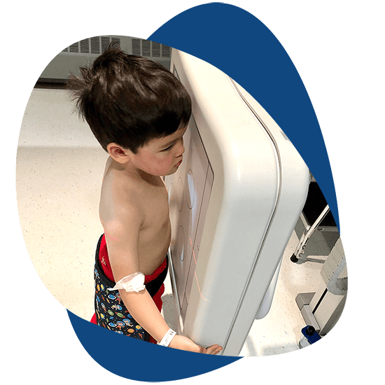
On March 26th 2018...
Four-year-old Liam Hugo was diagnosed with an extremely rare and life-threatening autoimmune disease called Juvenile Myositis (JM). Our once active son had deteriorated dramatically within a few short months.
-
![]()
When Liam Began...
Aggressive treatment for JM, he was unable to sit up, climb stairs, dress himself, or lift his own head without being in pain. Currently Juvenile Myositis has no known cause, no FDA approved treatments and no cure.
We’re on a mission to change that!
-
![]()
Although Liam's...
Disease does not define him, it’s important that we share his journey to help raise awareness for undiagnosed children, plus form bonds within our global Juvenile Myositis family.
-
![]()
Liam has...
An incredible imagination. He enjoys family fantasy story-time, playing pirates with his little sister Olivia and LOVES video games like Minecraft and ROBLOX. Liam is also quick to make friends, has excellent manners and tells fantastic jokes.
“What do you call a cow with no legs? GROUND BEEF!”
-
![]()
Follow Our Journey
With your help to spread JM awareness, there is more opportunity for quicker diagnoses, better treatment options, and ultimately finding a cure.
Together we can CureJM4Liam.


Our Journey


What is JM?
Juvenile Myositis (JM) and it’s two sub-forms, Juvenile Dermatomyositis (JDM) and Juvenile Polymyositis (JPM), are rare and life-threatening autoimmune diseases in which the body’s immune system attacks its own cells and tissues causing muscle weakness..
The exact occurrence of JM is unknown, however approximately 2 to 4 children in a million are diagnosed with JM each year in the United States. The average age of onset for JDM is between six to seven years old; 25% are age 4 or less.
-
The immune system is a group of cells that normally protects the body from infection and other environmental factors. Researchers believe a combination of environmental and genetic factors may influence the cause of JM.
Genetically, children who develop this disease often have a family history of other autoimmune diseases, such as thyroid disorder, type I diabetes, rheumatoid arthritis, lupus, or Crohn’s disease. If a child is genetically predisposed to JM, experts suspect a microbe, such as a virus or bacteria, might trigger a runaway immune response causing the immune system to attack previously healthy tissues, instead of protecting them.
Other environmental factors, such as a heavy dose of sun exposure, may also play a role.
-
WEAK MUSCLES
The muscles that are affected the most are near the trunk of the body: the stomach, thigh muscles, neck and upper arm muscles. Other muscles, however, can become weak as well. Sometimes there is inflammation in the swallowing tube or esophagus, which makes it difficult for the child to swallow. Other times, there is inflammation in the intestinal tract, which causes bowel difficulties and stomach pain.
A child with JM has great difficulty climbing stairs and just getting up from a sitting position. Walking and running become very challenging and exhausting. Some children will literally roll out of bed, because sitting up and lifting the head becomes too difficult. In addition to weakness, some children with JM experience pain in their muscles.
SKIN RASH
The skin rash in JDM usually occurs on the eyelids, knuckles, elbows, knees and ankles. The rash may appear before, after or at the same time as the muscle weakness. Sometimes the rash is so faint that it is not noticeable.
The rash appearing on the eyelids is red and purplish in color and is referred to as a heliotrope rash (named after a flower of the same color). Another rash that frequently occurs on the cheeks takes on a red color and looks very similar to a sunburn.
On the knuckles, elbows and knees, the rash takes the form of patches of red and scaly skin (called Gottron’s papules). The fingernails and the nail beds may take on a pinkish color as well.
FATIGUE
With JM, the child becomes tired easily and can only walk short distances. The child needs to rest often and lacks the energy for normal activities. It becomes difficult for the child to keep up with friends.
FEVER
Sometimes, the child with JM runs low-grade fevers, especially at night.
-
The following symptoms of JM are less common, but important to understand:
CALCINOSIS
Researchers believe approximately 10% to 50% of the children with JM will develop calcinosis. Calcinosis is the development of small lumps or linear deposits of calcium under the skin or in the muscle. They may feel like rocks under the skin and can range in size from a small pebble to a large softball. They can also form in a sheet-like appearance.
In many cases, these calcium deposits can grow over time or remain unchanged for years. In some cases, the calcinosis lumps are absorbed back into the body. Other times, the lumps break through the skin where they may leak creamy white calcium. These protrusions can become infected and painful, and sometimes need to be surgically removed. Calcinosis lumps are most likely to break through on the joints, such as the elbows and knees, but they can emerge anywhere on the body, often in pressure points.
DIGESTIVE TRACT COMPLICATIONS
The Gastrointestinal (GI) tract refers to the digestive system including the esophagus (swallowing tube), stomach and intestines. Myositis can cause the muscles in the GI tract to become inflamed and they may not be strong enough to move food through the digestive tract. It is important to immediately alert your medical team if your child has any GI tract complications.
Because the major problem in JDM is a systematic vasculopathy, which means that small blood vessels are damaged, the absorption of medication and nutrition from the gastrointestinal tract may be impaired. Doctors may sometimes offer alternative treatments, such as IV medications, IV nutrition or a feeding tube, to ensure that the medication and nutrition is properly absorbed. In some cases, a nutritionist or speech pathologist may also be consulted.
VASCULITIC ULCERS
Vasculitic ulcers are holes in the skin or gastrointestinal tract caused by inflammation of the blood vessels. When they occur in the skin, they look like open sores and can extend deep into the tissues. The ulcers can be very painful and generally are associated with a more severe course of JM.
When the ulcers occur in the gastrointestinal tract, they can harm the digestive organs and result in very serious illness. The warning signs of this condition are severe stomach pain, dark black stools or blood in the stools.
CONTRACTURES
Contractures are shortened muscles that cause a joint to stay in a bent position or have limited movement. Contractures can occur due to inflammation, muscle weakness, or calcinosis crossing over a joint.
LIPODYSTROPHY
Lipodystrophy is a loss of body fat. In areas affected by lipodystrophy, the fat cells become damaged, and if widespread and extensive, this can result in problems with storing fat and sugar, resulting in high cholesterol and diabetes.
-
A doctor will first perform a complete physical exam, specifically looking for rashes and muscle weakness. If JM is suspected, blood tests are the next step in making a diagnosis. Muscle enzymes are measured, including creatine kinase (CK), aldolase, lactate dehydrogenase (LDH), aspartate aminotransferase (AST), and alanine aminotransferase (ALT). If these lab tests are elevated, enzymes are leaking from inflamed or damaged muscles into the bloodstream. Antinuclear antibodies (ANA) are also measured to see if the child’s body is producing antibodies against its own cells. Several myositis-specific autoantibodies have also been identified, and these can be tested as well. Other blood tests are sometimes available to check out immune activation and/or blood vessel damage.
The next step in diagnosing JM is usually an MRI which can detect muscle inflammation and damage. If the MRI shows evidence of diseased muscles, a muscle biopsy may be performed to finalize the diagnosis. When the characteristic rashes are not present, it is important that a muscle biopsy be performed to exclude other causes of muscle weakness. A small amount of muscle is removed for examination under a microscope to determine if and how much the muscles and blood vessels have been affected by the disease.
If the patient has the rash and muscle weakness of JM, the lab tests are consistent with JM, and the MRI shows muscle inflammation, some doctors will not perform a muscle biopsy.
-
Although medications can help alleviate the symptoms of JM, the disease has no known cure. The primary medications used to treat the symptoms of JM are corticosteroids, immunosuppressants and chemotherapy. These medications themselves can cause severe side effects, making JM challenging to treat. Many researchers believe that early and aggressive treatment is usually the best predictor of a better outcome of this disease.
High dose oral Prednisone or other corticosteroids often coupled with intravenous corticosteroids (Solumedrol) are usually the first line of treatment for JM. Since the side effects of corticosteroids can be very troublesome, Methotrexate (a chemotherapy drug administered at much lower doses than used for cancer patients) is usually introduced early to allow for tapering of the corticosteroids.
Second-line treatments include Cyclosporine and Intravenous Immunoglobulin (IVIG). Plaquenil is often used to control the rash. For patients who do not respond to the first and second line of treatments, or in patients severely affected by the disease, Rituximab, Cytoxan and CellCept can be added. Protection from UVB is essential with sunscreens. Vitamin D with adequate calcium intake keeps bones strong. In addition to medications, physical therapy is generally prescribed to increase strength and flexibility in muscles and joints.
-
There is no cure for JM, but with advances in early diagnosis and aggressive treatment, the outcome has continued to improve. Some children experience a mild form of the disease and may go into remission. Others follow a more severe and potentially debilitating course that can be life-long. Some JM patients will have a loss of range of motion. Some will battle an array of serious complications, resulting in the inability to walk, ongoing pain, disfigurement and even death. Whether the course of the disease is mild or severe, JM is life-changing for all of these children and their families.
This website is provided for educational and informational purposes only. The information above was provided by the CureJM Foundation website.